Retina Vision

This is a blog on the information I feel to be relevant to the public/ health care providers and retinal patients with regards to eye care/ health esp where the retina is concerned.

Wednesday, October 3, 2012

Posterior Vitreous Detachment and Floaters!


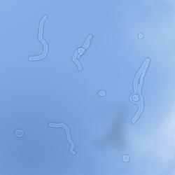
A gentleman walks into my clinic a few days back and says "You need to laser these black spots floating around in my eyes. They bother me so much, I can't work on the computer" he adds"I read you can laser them or go for a vitrectomy, I want the laser" I'm even willing to travel back to the US to get them lasered if you can't"..Floaters...Love em or hate em, they'll be there for you..


Problem: Floaters ( pesky flying irritants)
Treatment: nil. (YAG displacement  and Vitrectomies are a bit much considering the risks)
Who gets this? Everyone
When do you get it? At any time, usually more frequently with age
Should I worry? Read on....
The process of the vitreous(jelly)-at the rear of the eye, coming away from the retina is called a  “posterior vitreous detachment” or “PVD”

            As you age the vitreous becomes more watery, less jelly-like and isn't able to keep its usual shape. As a result, it begins to move away from the retina at the back of the eye towards the centre of the eye.
            A PVD is a natural change that occurs in the eye. Over 75 per cent of the population over the age of 65 develop a PVD, and it is not uncommon for it to develop in someone's 40s or 50s. PVD is not a sign of a disease or eye health problem. For most of us a PVD happens naturally as we get older.
PVD can cause symptoms such as floaters or little flashes of light across your vision. Floaters can take lots of different forms and shapes and can come in different sizes. You may see them as dots, circles, lines, clouds, or cobwebs. Sometimes, floaters can move around quickly. At other times it can feel like they hardly move at all. You may find floaters are more obvious in bright light or on a sunny day. The movement of the vitreous away from the retina at the back of the eye creates a tug on the retina. The retina reacts by sending a small electrical charge to your brain. You see this as short, small, flashes of light.

            Importantly, these same symptoms can be an indication of a more serious problem, such as a retinal tear, which needs urgent attention. You will not be able to tell the difference between floaters and flashes caused by PVD or retinal detachment. The only way you can tell is to have your eyes examined by an ophthalmologist.. If you suddenly experience any of the following symptoms, make sure you have your eyes examined as soon as possible - preferably on the same day or within 24 hours:
·    a sudden appearance of floaters or an increase in their size and number
·    flashes of light and/or a change/increase in the flashing lights you experience
·    blurring of vision
·    a dark 'curtain' moving up, down or across your vision, as this may mean that the retina has already partially detached.
           
            There is no medical treatment for PVD. There is no evidence to show that eye exercises, diet changes or vitamins can help a PVD. You may find floaters frustrating as they get in the way of seeing things which can make activities, such as reading, difficult. Sunglasses, dimming lights or UV eye shields limit the amount of light coming into your eye, which may help the floaters be less obvious, especially in bright conditions. However, most people find that over time the floaters become less of a problem and they don't need any special adaptations. Most people with a PVD have no restrictions on their activities. For more info: http://www.nei.nih.gov/health/vitreous/vitreous.asp

Monday, October 1, 2012

Do you want healthy eyes??


Eye Health Tips
Simple Tips for Healthy Eyes
Your eyes are an important part of your health. There are many things you can do to keep them healthy and make sure you are seeing your best. Follow these simple steps for maintaining healthy eyes well into your golden years.
Have a comprehensive dilated eye exam. You might think your vision is fine or that your eyes are healthy, but visiting your eye care professional for a comprehensive dilated eye exam is the only way to really be sure. When it comes to common vision problems, some people don’t realize they could see better with glasses or contact lenses. In addition, many common eye diseases such as glaucoma, diabetic eye disease and age-related macular degeneration often have no warning signs. A dilated eye exam is the only way to detect these diseases in their early stages.
During a comprehensive dilated eye exam, your eye care professional places drops in your eyes to dilate, or widen, the pupil to allow more light to enter the eye the same way an open door lets more light into a dark room. This enables your eye care professional to get a good look at the back of the eyes and examine them for any signs of damage or disease. Your eye care professional is the only one who can determine if your eyes are healthy and if you’re seeing your best.
Make sure to schedule eye check ups at the age of 40, 50, 55 and yearly then on.
Know your family’s eye health history. Talk to your family members about their eye health history. It’s important to know if anyone has been diagnosed with a disease or condition since many are hereditary. This will help to determine if you are at higher risk for developing an eye disease or condition. For example-glaucoma.
Eat right to protect your sight. You’ve heard carrots are good for your eyes. But eating a diet rich in fruits and vegetables, particularly dark leafy greens such as spinach, kale, or collard greens is important for keeping your eyes healthy, too. These contain high levels of Lutein and Zeaxanthine. The Chinese herb "keiji" or wolf berry is reputed to contain the highest levels of these products. Research has also shown there are eye health benefits from eating fish high in omega-3 fatty acids, such as salmon, tuna, and halibut.
Maintain a healthy weight. Being overweight or obese increases your risk of developing diabetes and other systemic conditions, which can lead to vision loss, such as diabetic eye disease or glaucoma. If you are having trouble maintaining a healthy weight, talk to your doctor. Exercising regularly keeps your weight in check and helps improve blood flow. It is said to reduce eye pressure by as much as 4 mmHg! 
Wear protective eyewear. Wear protective eyewear when playing sports or doing activities around the home. Protective eyewear includes safety glasses and goggles, safety shields, and eye guards specially designed to provide the correct protection for a certain activity. Most protective eyewear lenses are made of polycarbonate, which is 10 times stronger than other plastics. Many eye care providers sell protective eyewear, as do some sporting goods stores.
Quit smoking or never start. Smoking is as bad for your eyes as it is for the rest of your body. Research has linked smoking to an increased risk of developing age-related macular degeneration, cataract, and optic nerve damage, all of which can lead to blindness. It also worsens pre existing conditions such as diabetic retinopathy and glaucoma
Be cool and wear your shades. Sunglasses are a great fashion accessory, but their most important job is to protect your eyes from the sun’s ultraviolet rays. When purchasing sunglasses, look for ones that block out 99 to 100 percent of both UV-A and UV-B radiation.
Give your eyes a rest. If you spend a lot of time at the computer or focusing on any one thing, you sometimes forget to blink and your eyes can get fatigued. Try the 20-20-20 rule: Every 20 minutes, look away about 20 feet in front of you for 20 seconds. This can help reduce eyestrain. Having an adequate sleep is also helpful for its reparative properties. 
Clean your hands and your contact lenses—properly. To avoid the risk of infection, always wash your hands thoroughly before putting in or taking out your contact lenses. Make sure to disinfect contact lenses as instructed and replace them as appropriate.
Practice workplace eye safety. Employers are required to provide a safe work environment. When protective eyewear is required as a part of your job, make a habit of wearing the appropriate type at all times and encourage your coworkers to do the same.
Check your vision from time to time. Close one eye and then check the vision of the other eye by looking at a distant object. Alternate. A lot of conditions are missed as people do not notice a change in one eye as we use both eyes concurrently to view most things. 

Sunday, September 30, 2012

Diabetic Retinopathy- A Primer


Diabetic retinopathy

How Can Diabetes Affects Your Eyes?
Diabetes is a disease that affects the body’s ability to produce and/or use insulin in amounts sufficient to control blood sugar levels. There are three types of diabetes: type 1, type 2 and gestational, which may develop when a woman is pregnant:
  • Type 1:  usually diagnosed in children and young adults and previously known as juvenile diabetes. The body does not produce insulin.
  • Type 2:  the most common form of diabetes. Either the body does not produce enough insulin or the body’s cells ignore the insulin.
  • Gestational: blood sugar levels (glucose) become elevated during pregnancy in women who have never had diabetes before. Gestational diabetes starts when the mother’s body is not able to make and use all the insulin it needs during pregnancy.
People with any type of diabetes can develop hyperglycemia, which is an excess of blood sugar, or serum glucose. Although glucose is a vital source of energy for the body’s cells, a chronic elevation of serum glucose causes damage throughout the body, including the small blood vessels in the eyes.
As a result, if you have diabetes you run the risk of developing diabetic retinopathy, in which damage occurs to the delicate blood vessels inside the retina at the back of the eye. You are also at increased risk for developing cataracts (clouding of the normally clear lens in the eye), or glaucoma (a disease that results in damage to the optic nerve). However, with an annual dilated eye exam and control of blood sugar levels, ninety percent of vision loss can be avoided.

Description: http://www.geteyesmart.org/eyesmart/images/trans.gif
What Are Diabetic Retinopathy Risk Factors?
Several factors can influence the development and severity of diabetic retinopathy, including:
  • Blood sugar levels. Controlling your blood sugar is the key risk factor that you can affect. Lower blood sugar levels can delay the onset and slow the progression of diabetic retinopathy.
  • Blood pressure. A major clinical trial demonstrated that effectively controlling blood pressure reduces the risk of retinopathy progression and visual acuity deterioration. High blood pressure damages your blood vessels, raising the chances for eye problems. Target blood pressure for most people with diabetes is less than 130/80 mmHg.
  • Duration of diabetes. The risk of developing diabetic retinopathy or having your disease progress increases over time. After 15 years, 80 percent of Type 1 patients will have diabetic retinopathy. After 19 years, up to 84 percent of patients with Type 2 diabetes will have diabetic retinopathy. 
  • Blood lipid levels (cholesterol and triglycerides). Elevated blood lipid levels can lead to greater accumulation of exudates, protein deposits that leak into the retina. This condition is associated with a higher risk of moderate visual loss.
  • Ethnicity. While diabetic retinopathy can happen to anyone with diabetes, certain ethnic groups are at higher risk because they are more likely to have diabetes. These include African Americans, Latinos and Native Americans.
  • Pregnancy. Being pregnant can cause changes to your eyes. If you have diabetes and become pregnant, your risk for diabetic retinopathy increases. If you already have diabetic retinopathy, it may progress. However, some studies have suggested that with treatment these changes are reversed after you give birth and that there is no increase in long-term progression of the disease.
What is Diabetic Retinopathy?
Diabetic retinopathy, the most common diabetic eye disease, is caused by changes in the blood vessels of the retina. There are two types of diabetic retinopathy:
  • background or nonproliferative diabetic retinopathy (NPDR); and
  • proliferative diabetic retinopathy (PDR).

Description: http://www.geteyesmart.org/eyesmart/images/trans.gif

1. Nonproliferative Diabetic Retinopathy (NPDR)
Nonproliferative diabetic retinopathy (NPDR) is the earliest stage of diabetic retinopathy. With this condition, damaged blood vessels in the retina begin to leak extra fluid and small amounts of blood into the eye. Sometimes, deposits of cholesterol or other fats from the blood may leak into the retina. 
NPDR can cause changes in the eye, including:
  • microaneurysms - small bulges in blood vessels of the retina that often leak fluid.
  • retinal hemorrhages - tiny spots of blood that leak into the retina (these spots alone are rarely responsible for any loss of vision).
  • hard exudates - deposits of cholesterol or other fats from the blood that have leaked into the retina.
Many people with diabetes have mild NPDR, which usually does not affect their vision. However, if their vision is affected, it is the result of macular edema and macular ischemia.


 Macular edema is swelling or thickening of the macula. It is caused by fluid leaking from the retina’s blood vessels. The macula, which is responsible for our clear, central vision, does not function properly when it is swollen. Macular edema is the most common cause of vision loss in diabetes. Vision loss may be mild to severe, but in many cases, your peripheral (side) vision remains. Laser treatment may help to stabilize vision.
  • Macular ischemia occurs when small blood vessels (capillaries) close. Your vision blurs because the macula no longer receives enough blood to work properly. Currently, there is no effective treatment for macular ischemia.

2. Proliferative Diabetic Retinopathy (PDR)
Proliferative diabetic retinopathy (PDR) occurs when abnormal blood vessels begin to grow on the surface of the retina or optic nerve. This is called neovascularization. 
 PDR mainly occurs when many of the blood vessels in the retina close, preventing enough blood flow. The retina responds by growing new blood vessels in an attempt to supply the area where the original vessels closed. However, the new blood vessels are abnormal and do not supply the retina with normal blood flow. The new vessels are also often accompanied by scar tissue that may cause the retina to wrinkle or detach.
PDR may cause more severe vision loss than NPDR because it can affect both central and peripheral vision.
PDR causes vision loss in the following ways:
  • Vitreous Hemorrhage. The delicate new blood vessels may bleed into the vitreous—the gel in the center of the eye—preventing light rays from reaching the retina. If the vitreous hemorrhage is small, you may see a few new, dark floaters. A very large hemorrhage might block out all vision, allowing you to perceive only light and dark. Vitreous hemorrhage alone does not cause permanent vision loss. When the blood clears, your vision may return to its former level unless the macula has been damaged.
  • Traction Retinal Detachment. With PDR, scar tissue from neovascularization can shrink, causing the retina to wrinkle and pull from its normal position. This is called traction retinal detachment. Macular wrinkling can distort your vision. More severe vision loss can occur if the macula or large areas of the retina are detached.
  • Neovascular Glaucoma. Occasionally, if a number of retinal vessels are closed, neovascularization can occur in the iris (the colored part of the eye). In this condition, the new blood vessels may block the normal flow of fluid out of the eye. Pressure builds up in the eye, resulting in neovascular glaucoma, a particularly severe eye disease that causes damage to the optic nerve.
Diabetes can cause vision in both eyes to change, even if you do not have retinopathy. Rapid changes in your blood sugar alter the shape of your eye’s lens, and the image on the retina will become out of focus. After your blood sugar stabilizes, the image will be back in focus. You can reduce episodes of blurred vision by maintaining good control of your blood sugar.


Diagnosing Diabetic Retinopathy
The only way to detect diabetic retinopathy and to monitor its progression is through a comprehensive eye exam. There are several parts to the exam:
  • Visual acuity test. This uses an eye chart to measure how well you can distinguish object details and shape at various distances. Perfect visual acuity is 20/20 or better. Legal blindness is defined as worse than or equal to 20/200.
  • Slit-lamp exam.  A type of microscope is used to examine the front part of the eye, including the eyelids, conjunctiva, sclera, cornea, iris, anterior chamber, lens, and also parts of the retina and optic nerve.
  • Dilated exam. Drops are placed in your eyes to widen, or dilate, the pupil, enabling your Eye M.D. to examine more thoroughly the retina and optic nerve for signs of damage.
It is important that your blood sugar be consistently controlled for several days when you see your eye doctor for a routine exam. If your blood sugar is uneven, causing a change in your eye’s focusing power, it will interfere with the measurements your doctor needs to make when prescribing new eyeglasses. Glasses that work well when your blood sugar is out of control will not work well when your blood sugar level is stable.
In addition to a visual acuity test and a dilated exam, your Eye M.D. may find additional tests useful.

Description: http://www.geteyesmart.org/eyesmart/images/trans.gif
Fluorescein Angiography/Optical Coherence Tomography (OCT)
Your doctor may order fluorescein angiography or optical coherence tomography (OCT) to further evaluate your retina or to guide laser treatment if it is necessary. Fluorescein angiography is a diagnostic procedure that uses a special camera to take a series of photographs of the retina after a small amount of yellow dye (fluorescein) is injected into a vein in your arm. The photographs of fluorescein dye traveling throughout the retinal vessels show:
  • which blood vessels are leaking fluid;
  • how much fluid is leaking;
  • how many blood vessels are closed;
  • whether neovascularization is beginning.
OCT is a non-invasive scanning laser that provides high-resolution images of the retina, demonstrating its thickness. OCT can provide additional information regarding the presence and severity of macular edema.
These tests help the doctor determine:
  • why vision is blurred;
  • whether laser treatment should be started;
  • where to apply laser treatment.

Ultrasound
If your ophthalmologist cannot see the retina because of vitreous hemorrhage, an ultrasound test may be done in the office. The ultrasound can “see” through the blood to determine if your retina has detached. If there is detachment near the macula, this often calls for prompt surgery.
When the evaluation is complete, your ophthalmologist will decide when you need to be treated or re-examined.
People with diabetes should see their ophthalmologist right away if they have visual changes that:
  • affect only one eye;
  • last more than a few days;
  • are not associated with a change in blood sugar.

How Diabetic Retinopathy Is Treated
The best treatment is to prevent the development of retinopathy. Strict control of your blood sugar will significantly reduce the long-term risk of vision loss. Treatment will not usually cure diabetic retinopathy or restore normal visual acuity, but it may slow the progression of visual loss. Without treatment, diabetic retinopathy progresses steadily from minimal to severe stages.

Description: http://www.geteyesmart.org/eyesmart/images/trans.gif
Description: http://www.geteyesmart.org/eyesmart/images/break_dotted.jpg
Laser SurgeryLaser Surgery
The laser is a very bright, finely focused light. It passes through the clear cornea, lens and vitreous without affecting them in any way. Laser surgery shrinks abnormal new vessels and reduces macular swelling. Treatment is often recommended for people with macular edema, proliferative diabetic retinopathy (PDR) and neovascular glaucoma.
Laser surgery is usually performed in an office setting. For comfort during the procedure, an anesthetic eyedrop is often all that is necessary, although an anesthetic injection is sometimes given next to the eye. The patient sits at an instrument called a slit-lamp microscope. A contact lens is temporarily placed on the eye in order to focus the laser light on the retina with pinpoint accuracy.
For macular edema, the laser is applied near the macula in order to reduce fluid leakage. The main goal of treatment is to prevent further loss of vision by reducing the swelling of the macula. It is uncommon for people who have blurred vision from macular edema to recover normal vision, although some will experience partial improvement.
A few people may see laser spots near the center of their vision following treatment. They usually fade with time, but may not disappear completely.
In PDR, the laser is applied to all parts of the retina except the macula (called PRP, or panretinal photocoagulation). This treatment causes abnormal new vessels to shrink and often prevents them from growing in the future. It also decreases the chance that vitreous bleeding or retinal distortion will occur. Panretinal laser has proven to be very effective for preventing severe vision loss from vitreous hemorrhage and traction retinal detachment.


Side effects of panretinal laser surgery may include:
  • temporary blurred vision for days to a few weeks;
  • occasional mild loss of central vision;
  • mild to moderate loss of peripheral vision;
  • decreased night vision.
Multiple laser treatments over time may be necessary. Laser surgery does not cure diabetic retinopathy and does not always prevent further loss of vision. In spite of laser surgery, some people with PDR develop macular distortion or bleeding into the vitreous, causing blurred vision. Vitreous hemorrhage may recur and blood may accumulate faster than the eye can reabsorb it.
Vitrectomy Surgery
Vitrectomy is a surgical procedure performed in a hospital or ambulatory surgery center operating room. It is often performed on an outpatient basis or with a short hospital stay. Either a local or general anesthetic may be used.
During vitrectomy surgery, an operating microscope and small surgical instruments are used to remove blood and scar tissue that accompany abnormal vessels in the eye. Removing the vitreous hemorrhage allows light rays to focus on the retina again.
Vitrectomy often prevents further vitreous hemorrhage by removing the abnormal vessels that caused the bleeding. Removal of the scar tissue helps the retina return to its normal location. Laser surgery may be performed during vitrectomy surgery. Vitrectomy surgery may be recommended for:
  • thick vitreous blood in young, insulin-dependent diabetic patients. This is because they are at high risk for traction detachment, which may be hidden behind the blood.
  • thick or repetitive vitreous hemorrhage that does not clear or significantly interferes with the person’s lifestyle.
  • recent traction detachment or distortion of the macula.
To help the retina heal in place, your ophthalmologist may place a gas bubble in the vitreous space. The gas bubble will dissolve in one to eight weeks, depending on the type of gas used. You may be told to keep your head in certain positions while the gas bubble helps to heal the retina. It is important to follow your ophthalmologist’s instructions so your eye will heal properly. You will be advised not to travel by plane or to a high altitude while this bubble is in your eye, since a rapid increase in altitude can raise your eye pressure to dangerous levels and could cause vision loss or even blindness.
Risks of vitrectomy include infection, bleeding, retinal detachment and high pressure in the eye. Cataract formation is uncommon right after surgery; however, most patients often develop a cataract after a number of months or within a few years.


Advances in treatment
Researchers are studying treatments that target the underlying biochemical mechanisms that cause diabetic retinopathy. One  treatment involves the use of VEGF inhibitors, which hinder the growth of new blood vessels. Because leakage from these abnormal blood vessels causes vision loss, preventing their growth could avoid damage to the retina.
There is now widespread information on this treatment be it for DME or for Proliferative retinopathy. It is a great adjunct to our armamentarium. 
On the surgical front we have advanced our techniques and machinery. MicroincisionalVitrectomy Surgery (MIVS) has revolutionized the process making it safer and quicker. The high cut rates of modern vitrectors and its design allows surgeons to get close to the retina enabling perfect dissections. Healing is quicker and patients are more satisfied. 
With the advent of Ocriplasmin, there will be further potential in innovation as a separation of the posterior hyaloid is paramount in preventing the severe complication of tractional retinal detachments in patients. 


When to Schedule an Eye Examination
Diabetic retinopathy usually takes years to develop, which is why it is important to have regular eye exams. Because people with Type 2 diabetes may have been living with the disease for some time before they are diagnosed, it is important that they see an Eye M.D. without delay. The American Academy of Ophthalmology recommends the following schedule for people with diabetes:
  • Type 1: Within five years of being diagnosed and then yearly.
  • Type 2: At the time of diabetes diagnosis and then yearly.
  • During pregnancy: Pregnant women with diabetes should schedule an appointment with their ophthalmologist in the first trimester because retinopathy can progress quickly during pregnancy.